.jpg)
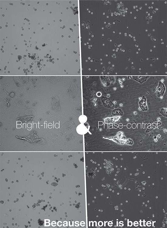
Thanks to the option to view the images either in clear field and in contrast, the evalution module receives more information and therefore is able to provide more accurate results.
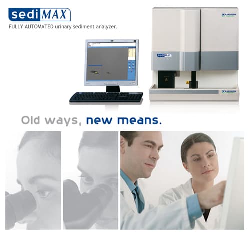
The urine sediment analysis next level
The optical system of the sediMAX conTRUST is capable of capturing images ofboth bright-field and phase-contrast microscopy to gain more information about the particles in the sample.
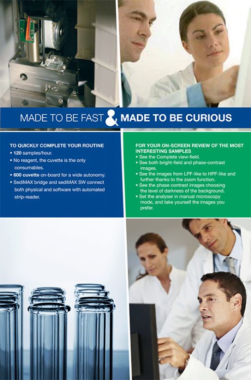
- Modified magnification. The images of the sediMAX conTRUST are LPF-like. (Low Power Field, which means a lower magnification and a larger field of view as compared to the sediMAX).
- Improved zoom function. An improved zoom function is implemented into the SW to better visualize details of particles. The operator can see HPF-like (as in case of sediMAX) or further magnified images
- Increased amount of investigated native sample. Due to the LPF-like whole viewfield images sediMAX conTRUST investigates 3x asmuch native urine per sample with the same throughput.
- Improved recognition (Both visual and automatic). Phase contrast microscopy converts phase shifts in light passing through a transparent specimen to brightness changes in the image, thus highlighting the areas where there is phase shift in the view field. This can help the detection of: Hyaline casts Ghost red blood cells, Squamous epithelial cells, Acanthocyte and yeast differentiation, Separation of cocci and rod bacteria, More clearly defined crystals.
- Composite image. The SW generates a composite image out of the bright-field and phase contrast microscopy images to show the features of each image in one view.
- Quantitative RBC and WBC evaluation. Due to the improvements of the measurement technique and the evaluation module, RBC and WBC evaluation is now quantitative.
- New way of bacteria evaluation. The evaluation module now recognizes cocci and rod bacteria separately.
- Manual microscopy mode. The instrument performs sample identification, mixing, aspiration into the cuvette and centrifugration. After the cuvette arrives at the microscope the user has the option to move the cuvette via on-screen buttons and take images of any area of the cuvette, changing focusing height. During the process the user has a live view of the cuvette. Images taken arethen evaluated by the new evaluation module.
Sedimax can be connected to A.Menarini Diagnostics strip readers to form a fully automated system combining urine sediment and urine chemistry analysis.
| Sample Preparation | Automatic, on-board |
| Sample volume | 2ml |
| Analysis volume | 0.2ml (200ul) |
| Throughtput | Up to 120 samples/hr |
| Load capacity | 100 sample tubes (10 samples x 10 racks) |
| Sample tube spec | Ht 100 – 105 mm, Diam 16 – 17.5 mm |
| Barcode reader | On-board |
| SediMAX cuvette pack | 50 cuvettes in 12 towers (600 cuvettes in total) |
| Cuvette carousel capacity | 600 cuvettes (50 x 12 towers) |
| Centrifuge | On-board. 2000 rpm for 10 secs per sample |
| Automated microscopy analysis | 10, 15 & 20 high power fields (HPF) equivalent. Selectable. |
| Final result | Particle concentration (HPF or /ul) & identified category |
| Detected particles | RBC, WBC, Hyaline casts, Pathological casts, Epithelial cells, Small round cells, Bacteria, Yeasts, Crystals: Calcium oxalatemonohydrate, Calcium oxalate dihydrate, Uric acid,Tri-phosphate, Mucus, Sperm |
| Resuls archieve | 10.000 sample results and images |
| Wash solution | Distilled water (5L bottle), hypochlorite/ bleach solution |
| Waste bin capacity | 500 cuvettes |
| Size & weight | 600 x 600 x 600 (h,d,w). 58kg |
Downloads
- sedimax